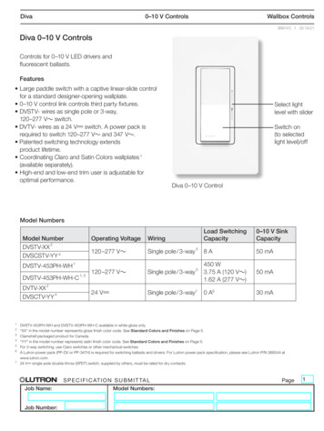
Transcription
Examiningthe FingernailsMark E. Williams, M.D.University of VirginiaSchool of Medicine
PreTest 1 What disease wouldmost likely producethese nails?– Diabetes mellitus– Congestive heartfailure– Hypothyroidism– Liver disease– Renal disease– SLE
PreTest 2 What disease wouldmost likely producethese nails?– Diabetes mellitus– Congestive heartfailure– Hypothyroidism– Liver disease– Renal disease– SLE
PreTest 3 What disease wouldmost likely producethese nails?– Diabetes mellitus– Congestive heartfailure– Hypothyroidism– Liver disease– Renal disease– SLE
What information is availablefrom examining the fingernails? Overall vitalityInner emotional stateCerebral dominanceOccupations andhobbies Past medical history Nutritional status Cardiovascularfunction Rheumatic conditions Dermatologicalproblems
Sequence of the Examination Check the nail shapeExamine the nail colorSurvey processes around the nailsCompare handsNote skin conditions
Observing the Nail Shape
Normal Nail Findings Softness and flexibility offree edgeShape and colorQuality of paronychial tissueGrowth rate –– Six months from cuticle tofree edgeTime of events can beestimated from locationNail polish––Distance from base and lineof polish gives approximatedate of application (nailsgrow 0.1mm/ dayToenail polish suggestsunusual flexibility, a friendlyhelper, or pedicure
Clubbed fingernails Causes of clubbing (not exhaustive)– Pulmonary and Cardiovascular causes(80%) Lung cancer, pulmonic abscess,interstitial pulmonary fibrosis,sarcoidosis, beryllium poisoning,pulmonary arteriovenous fistula,subacute bacterial endocarditis, infectedarterial grafts, aortic aneurysm– Gastrointestinal causes (about 5%) Inflammatory bowel disease, sprue,neoplasms (esophagus, liver, bowel)– Hyperthyroidism (about 1%)– Note: Chronic Obstructive PulmonaryDisease does not cause clubbing!Image courtesy
Schamroth’s signPurpose- to determine if nails areclubbedMethod-have patient place bothforefinger nails togetherand look between them.If you can see a smalldiamond space betweenthem (Schamroth’swindow) then the nailsare not clubbed
Spooned nails (Koilonychia) Water drop test– Imagine placing a drop ofwater on the nail with amedicine dropper. If a drop ofwater would not roll off thenail, it is spooned Causes– iron deficiency– diabetes mellitus– Protein deficiency especially insulfur-containing amino acids(cysteine or methionine)Koilonchychia comes from the Greek words for “spoon” and “nail”.
Beau’s Lines Caused by growtharrest Sign of significantillness Temporalrelationships (locationof the line tells whenthe illness wasexperienced)The location half way up the nailSuggests illness 3 months agoNote the 2 Beau’s linesAbout 2 months apart
Thin Brittle Nails Metabolic bone disease– Nail thinness is correlatedwith osteopenia Thyroid disorder Systemic amyloidosis– Yellow waxy flaking Severe malnutritionNote the thin nails in this womanwith severe osteopeniaSystemic Amyloidosis
Central Nail Ridge Causes– Iron deficiency– Folic acid deficiency– Protein deficiency
Central Nail Canal(Median Nail Dystrophy) “Heller’s fir treedeformity” Cuticle is usuallynormal Associations– Severe arterialdisease– Severe malnutrition– Repetitive trauma
Nail Pitting Cause is nail matrixinflammation Conditions– Psoriasis (randomappearance of pits)– Alopecia areata(geometric rippledgrid)– Eczema– Lichen planusImages courtesy
Nail Beading Beads seem to dripdown the nail likewax Associated withendocrine conditions– Diabetes mellitus– Thyroid disorders– Addison’s diseaseImage courtesy
Rough Nail Surface Nails look sandpaperedand dull Consider:––––autoimmune diseasePsoriasisChemical exposureLichen planus
Nail Thickening Slow nail growthproduces thethickness Consider:– Onychomycosis– Chronic eczema– Peripheral vasculardisease– Yellow nail syndrome– Psoriasis
Separation of the Nail Plate(Onycholysis) Caused by lifting of thenail plate sisTraumaContact dermatitisToxic exposures (solvents)Porphyria cutanea tarda(onycholysis and blisteringof sun exposed skin)Traumatic onycholysis(Only involving one nail)PsoriasisNote the jagged border
Severe Curvature Curved or beakednails– Caused by resorptionof distal digit– Consider HyperparathyroidismRenal failurePsoriasisSystemic sclerosis
Complete Nail DestructionLocal mechanisms:– Trauma– ParonychiaGeneralized conditions:– Toxic epidermal necrolysis– Chemotherapy– Bullous diseases– Vasculitis
Observing Nail Color
Abnormalities of the Lunula Absent– Anemia– Malnutrition Pyramidal– Excessive manicure– Trauma Red Discoloration––––Cardiovascular diseaseCollagen vascular diseaseHematological malignancyOthers
Focal Discolorationsof the Nail Plate
Transverse White Lines Mee’s lines– Can time the event from locationon nail– Significant illness– Heavy metal toxicity– Chemotherapy Muehrcke’s lines––––Parallel white irregular linesCaused by edema to nail plateSign of hypoalbuminemiaLines do not migrate anddisappear when albumin increases
White Splotches Leukonychia striae Caused by minortrauma to the nailmatrix Timing can bedetermined by thelocation on the nail
Longitudinal Brown Lines Mechanism– Increased melaninproduction by nail matrixmelanocytes Associations––––Addison’s diseaseNevus at nail baseBreast cancerMelanoma (check forperiungal pigmentation)– TraumaImage courtesy
Splinter Hemorrhages Caused byhemorrhage of distalcapillary loops Note thickness yriasis rubra pilarisPsoriasisRenal failureSplinter hemorrhages tend to be fat.
Terry’s Half and Half Nails Proximal portion is white(edema and anemia) andthe distal portion is dark These nails imply eitherrenal or liver disease In renal disease there is abrown band at thejunction of the erythemaand the free edgeLiver disease (no brown line)Renal disease (brown line)Lower Image courtesy
Generalized Discolorationsof the Nail Plate
White Nails Caused by anemia,edema or vascularconditions Consider:––––––AnemiaRenal failureCirrhosisDiabetes mellitusChemotherapyHereditary (rare)
Pink or Red Nails Consider:– Polycythemia (dark)– SLE– Carbon monoxide(cherry red)– Angioma– Malnutrition
Brown Grey Nails Consider:– Cardiovasculardisease– Diabetes mellitus– Vitamin B12 deficiency– Breast cancer– Malignant melanoma– Lichen planus– Syphilis– Topical agents
Yellow Nails Consider:– Amyloidosis– Lymphedema andbronchiectasis (yellownail syndrome)– Median/Ulnar nerveinjury– Thermal injury– Jaundice– Diabetes mellitus
Green or Black Nails Topical preparations Chronic Pseudomonasinfection Trauma
Processes Around the Nail
Processes Around the Nail Chronic paronychialinflammation– Swelling– Scaling– Nail separation
Periungal telangeictasia Dilated capillary loopsand atrophy of cuticle Strongly associatedwith collagen vasculardisease– SLE– Dermatomyositis– SclerodermaImage courtesy
Swelling Around the Nail Mucus cyst Fibroma Malignantmelanoma
Masses Pyogenic granuloma Warts Fibroma Malignant melanoma
PreTest 1 What disease wouldmost likely producethese nails?– Diabetes mellitus– Congestive heartfailure– Hypothyroidism– Liver disease– Renal disease– SLE
PreTest 1 What disease wouldmost likely producethese nails?– Diabetes mellitus– Yellow nails andlongitudinal ridgingDiabetes Mellitus
PreTest 2 What disease wouldmost likely producethese nails?– Diabetes mellitus– Congestive heartfailure– Hypothyroidism– Liver disease– Renal disease– SLE
PreTest 2 What disease would mostlikely produce thesenails?– Terry’s half and half nailssuggest Liver disease or Renal disease– No convincing brown line atthe junction of theerythema and free edgesuggests liver diseaseLiver Disease
PreTest 3 What disease wouldmost likely producethese nails?– Diabetes mellitus– Congestive heartfailure– Hypothyroidism– Liver disease– Renal disease– SLE
PreTest 3 What disease wouldmost likely producethese nails?– SLE– Note the fat splinterhemorrhage and theperiungaltelangeictasiaSLE
Summary Considerable useful information isavailable from careful examination of thenails Starting with the nail examinationimmediately communicates a sense ofdiligence and thoroughness Remain attentive and continually add toyour diagnostic repertoire
Post Test 1 71 year old man withlethargy, fatigue, andanorexia What is yourdiagnosis and theevidence thatsupports it?
Renal Failure withhyperparathyroidism Terry’s half and halfnails imply liver orrenal disease Brown distal colorationsuggests renal disease Nail curvature impliesresorption of distalphalange from PTH
Post Test 2 66 year man withfatigue,hypotension andan increasedsense of smell
Addison’s disease Note longitudinal darkbrown line andbeading No cuticular nevus ormass to suggestmelanoma
Post Test 3 78 year old withdiabetes mellitus,anemia, congestiveheart failure andperipheral vascularinsufficiency What is the evidence?
Post Test 3 Red lunula can implyCHF Heller’s line suggestsperipheral vasculardisease Ridging suggestsdiabetes or anotherendocrine condition Overall pallorsuggests anemia
Post Test 4 84 year old man witha painful ankle. Name5 likely diseases onhis problem list
Post Test 4 Gout (tophi)CHF (red lunula)Anemia (pallor)Peripheral arterialdisease (longitudinalred line) Chronic kidneydisease (distal brownpigmentation)
Post Test 5 68 year old man withweight loss anddysphagia
Esophageal Cancer Extreme nail beakingimplies resorption ofdistal digit In this case due toPTH like hormoneproduced by themalignancy Loss of lunula impliesmalnutrition oranemia
Post Test 6 62 year old womanwith proximal muscleweakness, dysphagiaand weight lossImage courtesy
Dermatomyositis Cuticular atrophy andperiungaltelangiectasiassuggest collagenvascular disease Gottron’s papulesover the knucklesimplydermatomyositisImage courtesySystemic lupus affecting the skin over the handstends to spare the knuckles whileDermatomyositis tends to involve the knuckles
Case 7 78 year old manwith first degreeheart block,periorbital purpura,and an enlargedliver
Case 7Systemic Amyloidosis Thinned, raggededges Yellowdiscoloration Ridging
Case 8 70 year old man withdepression, fatigue,weight loss andirritability No history of trauma No evidence ofPsoriasis on exam
Case 8 Nails show significantonycholysis Pallor and loss oflunula suggestsmalnutritionThyrotoxicosis
Case 9 60 year old withpainful fingers
Case 9 Onycholysis Splitting of nailplate Salmon patch Chronicparonychia Nail disease isassociated withpsoriatic arthritisPsoriasis
Case 10 75 year old man withweight loss andshortness of breath
Case 10 Significant clubbing Nicotine staining fromcigarette smokingLung cancer
Case 11 Ill appearing 60year old man withfever, malaise,weight loss andpainful testicles
Case 11 Polyarteritis NodosaSplittingThinningRidgingNail plate infarctionPeriungal telangiectasia
Case 12 55 year old womanwith fatigue, musclepain and pleuriticchest pain
Case 12 Periungaltelangiectasis andcuticular atrophysuggest collagenvascular disease Rash tends to sparethe knuckleSystemic Lupus
Case 13 55 year old man withhyperpigmentation ofhis face, increasedfacial hair and darkurine
Case 13Porphyria CutaneaTarda Onycholysis andblistering Due to defective liveruroporphyrinogendecarboxylase Urine has coral pinkfluorescence under aWood's lamp
Acknowledgements The UVA GME Office for funding support Dr. Vladimir Goodkovski for technicalassistance Dr. Jim Thomas forpermission to use images from theirextensive dermatological atlas Internal Medicine residents at UVA for pretesting and helpful feedback